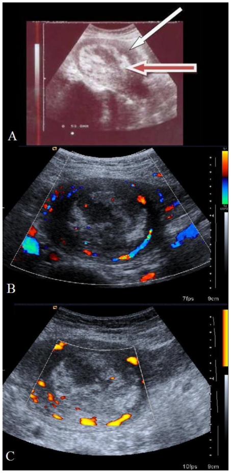
Figure 3.
28 year old primipara with placenta increta after methotrexate administration, 19th post-natal day - transabdominal sonography. 3A: Sagittal section (Siemens-Antares; 3.5MHz probe) showing increased echogenicity of placenta. Red arrow shows echogenic placental mass and white arrow points to the myometrium. 3B: Color Doppler (Siemens Antares; 3–5 MHz probe) showing decreased blood flow to the placenta (cross section). 3C: Power Doppler (Siemens Antares; 3–5 MHz probe) showing reduced blood flow to the echogenic placental mass

