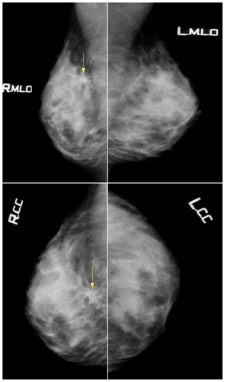
Figure 1.
47-year-old female patient with invasive lobular breast carcinoma metastatic to the urinary bladder. Mammography images (26 kV, 51mAs for MLO; 26 kV, 48 mAs for CC) in Craniocaudal and Mediolateral Oblique views, demonstrating: In the right breast, at the borderline of the upper quadrants, deep in the chest wall about 7cm from the areola and 4cm from the skin surface - a visible architectural distortion with spiculations radiating from the common center, measuring up to 30mm, with no associated mass and minor area of focal retraction. No microcalcifications were noted. Mammography of the left breast is unremarkable. This finding was later confirmed as Right Breast Invasive Lobular Carcinoma.

