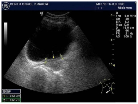
Figure 2.
47-year-old female patient with invasive lobular breast carcinoma metastatic to the urinary bladder. Trans-abdominal grayscale ultrasound image of urinary bladder (GE 4 MHz, Convex Transducer) in transverse plane, presenting: Irregular, isoechoic to bladder tissue, left segment urinary bladder wall thickening, involving left ureter outlet. This finding is consistent with breast cancer metastasis to the bladder.

