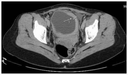
Figure 5.
47-year-old female patient with Invasive Lobular Breast Carcinoma. Contrast Enhanced Computed Tomography image in Arterial Phase. (GE LightSpeed 16-Slice Scanner; 290mAs, 120 kV, 5.0mm slice thickness, Ultravist intravenous contrast agent - dose 60ml administered at a rate of 3ml/sec). Axial section demonstrating: Hyperdense segmental urinary bladder wall thickening involving left lateral (white arrow), inferior and right lateral bladder wall. The contrast enhancement is 82jH (from 38jH before i.v contrast injection). There are no signs of involvement of adjacent structures (uterus and adnexa).

