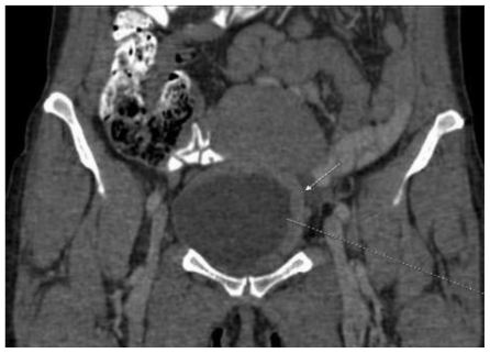
Figure 7.
47-year-old female patient with invasive lobular breast carcinoma metastatic to the urinary bladder. Contrast Enhanced Computed Tomography image of the pelvis in arterial phase (GE LightSpeed 16-Slice Scanner; 290 mAs, 120 kV, 1.2mm slice thickness, Ultravist intravenous contrast agent - dose 60ml administered at a rate of 3ml/sec). Coronal reconstruction demonstrating: Hyperdense segmental urinary bladder wall thickening up to 13mm, involving left lateral (white arrow) and superior bladder wall. There are no signs of adjacent structure involvement.

