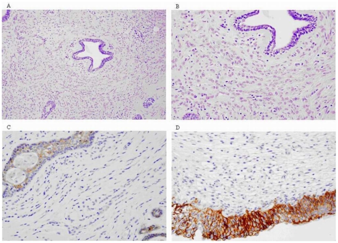
Figure 9.
47-year-old female patient with invasive lobular breast carcinoma metastatic to the urinary bladder. (A-C) Post-TURB Microscopic images presenting typical histological features of Breast Invasive Lobular Carcinoma: (A,B) - Single infiltrating tumour cells forming distinctive indian files and targetoid patterns (HE Stain; Magnification 100x and 200x respectively). (C) - Cancer cells’ lack of cohesion and E-cadherin expression (Immunohistochemical Stain; Magnification 200x). (D) Post-TURB Microscopic image presenting histological picture of metastatic lobular cancer to the urinary bladder: positive reaction to E-cadherin visible in the bladder epithelia only (Immunohistochemical Stain; Magnification 200x).

