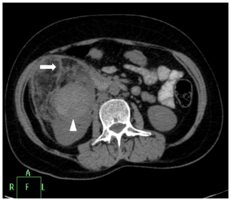
Figure 2a.
42 yr female with aneurysm formation in right renal angiomyolipoma: Plain axial CT scan (Philips 40 slice multidetector helical CT, at lumbar level) shows mixed density right renal mass with areas of fat attenuation (arrow). Few areas of hyperdensity were also noted within the mass (arrowhead). Parameters used: kVp - 120; mA - 306; slice thickness - 3mm.

