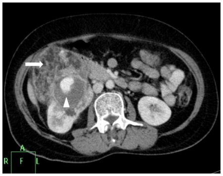
Figure 2b.
42 yr female with aneurysm formation in right renal angiomyolipoma: Contrast enhanced axial CT (Philips 40 slice multidetector helical CT at same level in arterial phase using non-ionic iodinated contrast agent Iohexol 90 ml manually injected) shows moderate heterogenous enhancement of the mass along with septal enhancement (arrow head). Intense vascular equivalent enhancing area is seen within mass consistent with partially thrombosed aneurysm (arrow). Parameters used: kVp - 120; mA - 206; slice thickness - 2mm.

