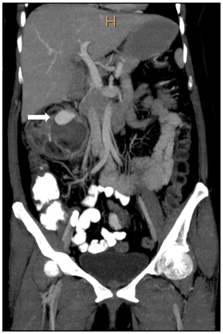
Figure 2c.
42 yr female with aneurysm formation in right renal angiomyolipoma: Coronal reformatted CT in maximum intensity projection (Philips 40 slice multidetector helical CT in arterial phase using non-ionic iodinated contrast agent Iohexol 90 ml manually injected) clearly demonstrates fatty right lower pole renal mass with partially thrombosed aneurysm (arrow). Parameters used: Parameters used: kVp - 120; mA - 206; Maximum Intensity Projection applied.

