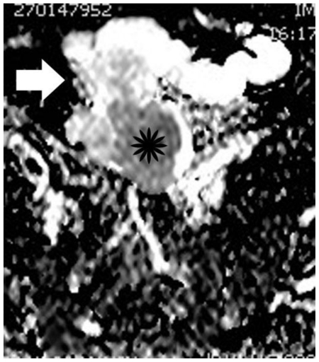
Figure 10.
30-year old woman with endometrioid adenocarcinoma developed in a pre-existing ovarian endometrioma. Apparent diffusion coefficient map of image illustrated in Figure 9. The soft tissue component (asterisk) is hypointense, with an ADC value of 1.05 × 10⁻³ mm2/s, a finding suggestive of restricted diffusion. Subacute hemorrhagic component (arrow) is detected with intermediate signal intensity and an ADC value of 1.37 × 10⁻³ mm2/s. The normal ADC values of myometrium and endometrium in this case were 1.79 × 10⁻³ mm2/sec; and 1.43 × 10⁻³ mm2/sec, respectively.

