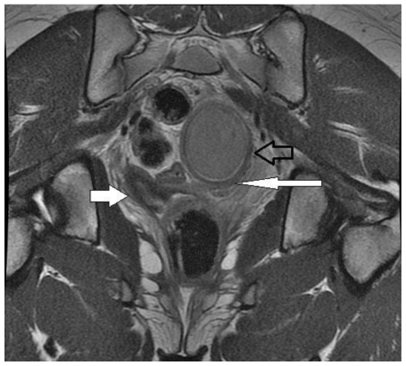
Figure 4.
13 year old female with uterus didelphys, unilateral distal vaginal agenesis, and ipsilateral renal agenesis. Coronal long axis non-fat saturated FSE T2. Left uterine horn and distended, blind-ending left lower uterine segment ended in intermediate signal tissue at the superior aspect of what appeared to be the vagina as noted on prior MRI (open arrow). Normal right uterine horn (filled arrow) and blind-ending uterus (long arrow). (Protocol: 1.5 Tesla MRI (GE Signa Excite), TR/TE: 4000/110, 5mm slice thickness, non-contrast).

