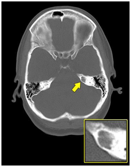
Figure 1.
A 48-year-old female with a left-sided intraosseous schwannoma of the petrous apex measuring 8.5 mm in the anteroposterior, 7 mm in the transverse, and 7 mm in the craniocaudal dimensions. Axial plain CT scan of the brain at the level of the petrous apex demonstrates a well-defined, smoothly marginated, lytic lesion with no internal matrix and no aggressive features located in the left petrous apex (arrow). (Protocol: 420 mAs, 140 kVp, 4.8 mm slice thickness).

