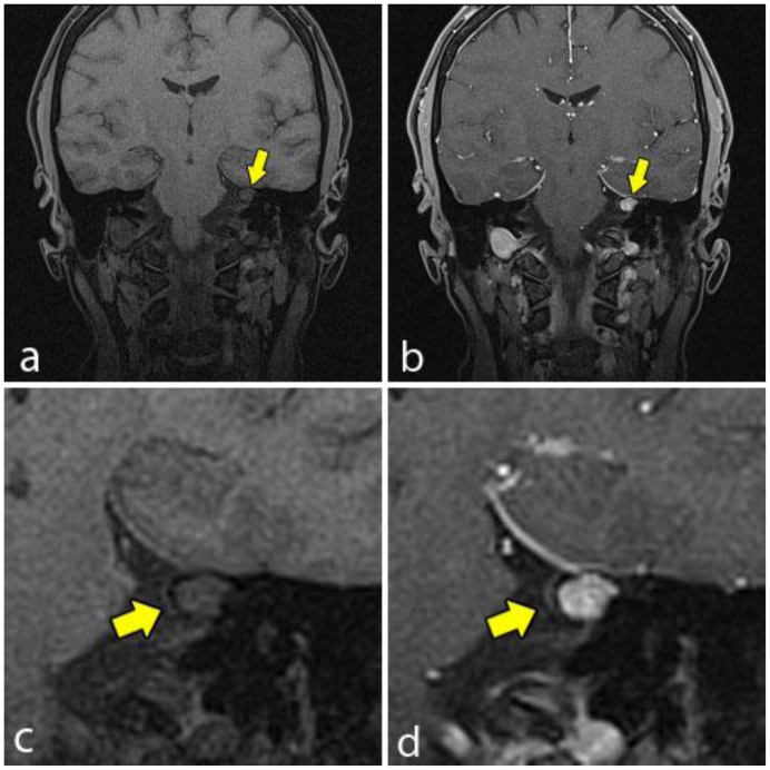
Figure 4.
A 48-year-old female with a left-sided intraosseous schwannoma of the petrous apex measuring 8.5 mm in the anteroposterior, 7 mm in the transverse, and 7 mm in the craniocaudal dimensions. Coronal T1W high resolution fat saturation MRI pre (4a, 4c) and post (4b, 4d) gadolinium administration. The pre contrast image (4a, 4c) demonstrates the left petrous apex lesion (arrow), which is isointense to brain and demonstrates no involvement of the cranial nerves. The post contrast image (4b, 4d) demonstrates solid enhancement of the same lesion. (Protocol: 4a, 4c; Magnet strength 1.5 Tesla, TR 5.37, TE 2.54, 1 mm slice thickness. 4b, 4d; Magnet strength 1.5 Tesla, TR 5.37, TE 2.54, with 14 mL of Multihance gadolinium, 1 mm slice thickness).

