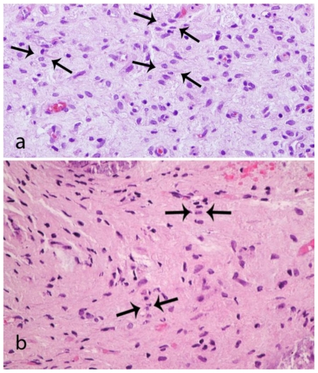
Figure 6.
A 48-year-old female with a left-sided intraosseous schwannoma of the petrous apex measuring 8.5 mm in the anteroposterior, 7 mm in the transverse, and 7 mm in the craniocaudal dimensions. Composite photomicrograph of two tissue sections (6a and 6b) demonstrating nuclei arranged in palisades (arrows). This is consistent with Antoni A tissue which is a characteristic feature of schwannomas. (Hematoxylin and eosin, original magnification 400x)

