Figure 2.
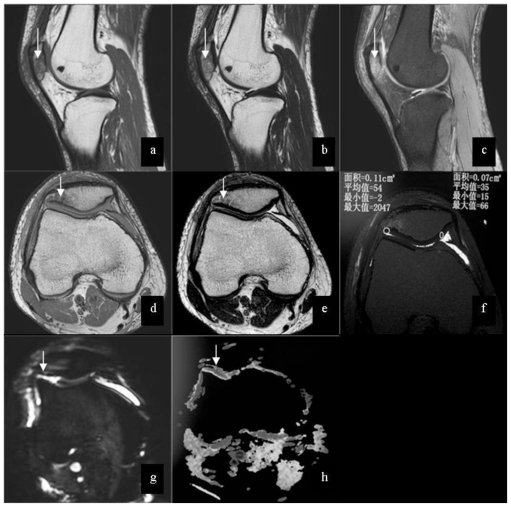
25-year-old male patient with injury of right knee. Images shows low signal (arrow) on T1WI (3.0 Tesla, TR/TE = 2165 msec/ 30msec, Slice Thickness = 3mm) (a), mixed signal (arrow) on T2WI(3.0 Tesla, TR/TE = 2231 msec/ 100msec, Slice Thickness = 3mm) (b) and high signal(arrow) on PDW-SPAIR(3.0 Tesla, TR/TE = 415msec/ 8msec, Slice Thickness =3 mm) (c) in the bone marrow of patella. The axial images show low signal (arrow) on T1WI (d) and mixed signal (arrow) on T2WI (e) in the bone marrow of patella, and no abnormal signal in the cartilage of patellar is shown. On T2 map (f) (3.0 Tesla, TR/TE=2400 msec/15, 30,45,60,75,90msec, Slice Thickness = 3mm), the mean value of the right part of cartilage is 54, and the mean value of the left part of cartilage is 35. There are slightly high signal (arrow) on DWI (g) (3.0 Tesla, TR/TE=2000 msec/70 msec, slice thickness=3 mm, b =600 s/mm2 ) and high signal (arrow) on ADC(h) compared with normal cartilage.

