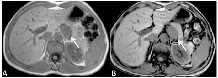
Figure 4.
35-year-old women with solid pseudopappilary tumor. Axial T1 weighted MR image in-phase (A) and out-of-phase (B) demonstrates a well defined, hypointense lesion adjacent to the posterior border of the pancreatic body-tail. No signal loss in out-of-phase images excluded the presence of microscopic fat content of the lesion. Fat cleavage plane with pancreatic gland is lost, but no signs of local invasion are identified. [Philips Intera 1.5 T, 5mm slice thickness, TE=171, TR=4,6 and TR=2,3]

