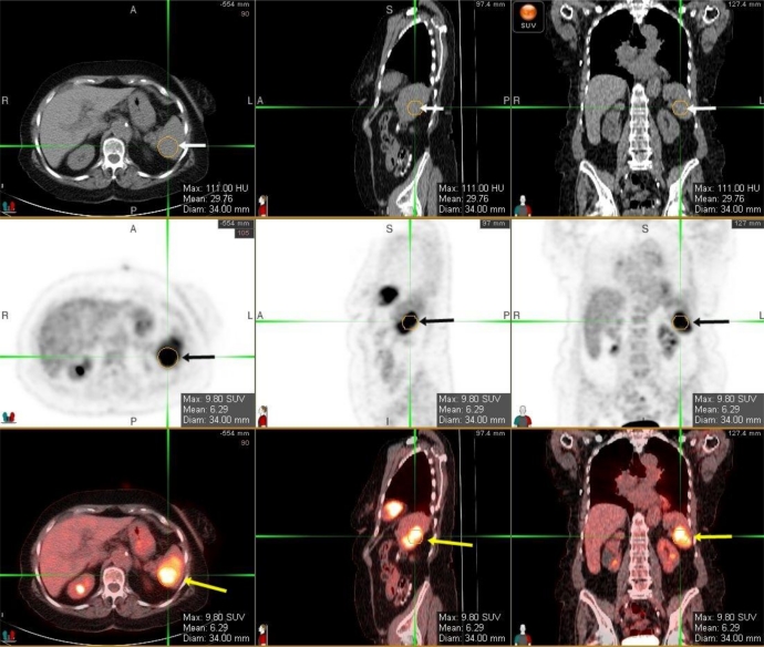
Figure 2.
Unenhanced CT scan (upper row) with PET scan images (middle row) and fused PET/CT scan (lower row images) of a splenic lesion, in a 91-year-old female with IPS. Images demonstrate focal area of increased radiotracer uptake in the spleen (black arrows on the PET scan and yellow arrows on the fusion images). This lesion corresponds with the area of low attenuation in the spleen on unenhanced CT scan (white arrows). The max SUV value of the lesion within the spleen is 9.8. (Philips GEMINI's PET/CT scanner; Performed 60 minutes after injection of 17.1 mCi FDG tracer, with 5 mm slice thickness, CT-based attenuation correction algorithm using two iterations and 8 subsets, 120 kVp, 250 mAs).

