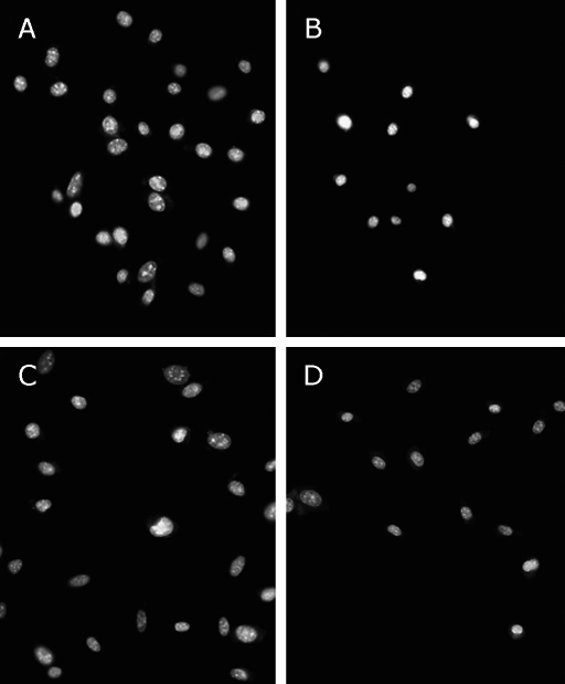
Fig. 3.
Effect of TFs on 6-OHDA-induced apoptotic nuclear morphology in SH-SY5Y cells. Fluorescence micrographs of SH-SY5Y cell nuclei from untreated cells (A); cells exposed to 100 µM 6-OHDA for 24 h (B); Cells pre-incubated with 0.5 µg/ml TFs (C) or 1 µg/ml TFs (D) for 3 h, and treated with 100 µM 6-OHDA for 24 h. The cells were stained with the DNA-binding fluorochrome Hoechst 33258. Scale bar = 50 µm.

