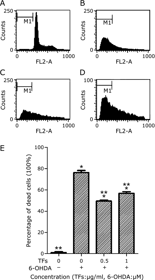
Fig. 4.
Cell apoptosis detected by flow cytometry. SH-SY5Y cells were incubated in drug-free medium (A) or medium containing with 100 µM 6-OHDA for 24 h (B); or cells were pre-incubated with 0.5 µg/ml TFs (C) or 1 µg/ml TFs (D) for 3 h. The results shown in (E) are the cell apoptosis rate of the means and SE for two independent experiments. The cells were stained with PI. Data are expressed as percentage of the dead cells means ± SD, n = 2. *p<0.01 significantly different from untreated control cells and **p<0.01 significantly different from 6-OHDA group.

