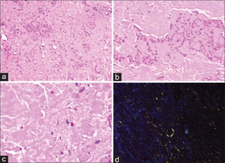
Figure 2.

Photomicrograph showing polygonal epithelial cells amidst amyloid-like material; note the absence of calcification (a) (H and E, ×100). Cells showing pseudoglandular pattern (b) (hematoxylin and eosin; ×200). Higher magnification of amyloid-like material (c) (H and E, ×400). Congo red staining showing green birefringence of amyloid (d) (polarizing microscop, ×200)
