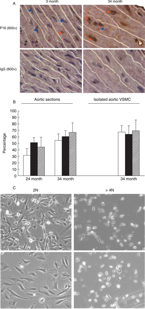
Fig. 1.
Polyploid vascular smooth muscle cells (VSMC) express p16INK4a and lose replication potential. (A) Aortic tissue sections derived from young and old Brown Norway rats were fixed with paraformaldehyde and subjected to immunostaining as detailed under the Supplementary material. The orange arrows point to p16INK4a-positive cells (brown staining localized to the nucleus) and the blue ones to p16INK4a-negative cells. Staining with anti-IgG was used as control. Shown are phase-contrast images at ×600 magnification, representative of three rats in each group. Staining was also carried out in cryosections, showing similar results (not shown). (B) The filled bars depict the percentage of polyploid cells identified based on nuclear size (for tissue sections), or 4′,6-diamindino-2-phenylindole (DAPI) staining (for isolated VSMC), the empty bars represent the percentage of polyploid cells that are also p16INK4a-positive, and the striped bars represent the percentage of p16INK4a-positive cells that are polyploid. Five slides per rat were analyzed, and a total of two or three rats were examined in each age group. Shown are average percentages ± standard deviations. (C) VSMC were isolated from 34-month-old aortas, and sorted by flow cytometer to obtain diploid (2N) and ≥ 4N populations, as described in Jones & Ravid (2004). Equal numbers of cells were cultured and monitored as in Nagata et al. (2005). Shown are phase-contrast microscopy images of the cells at 2 (lower panels) and 7 days after culturing. A small fraction of the polyploid population proliferated, but this also corresponds to the number of contaminating diploid cells in this pool (up to 10%).

