Fig. 2.
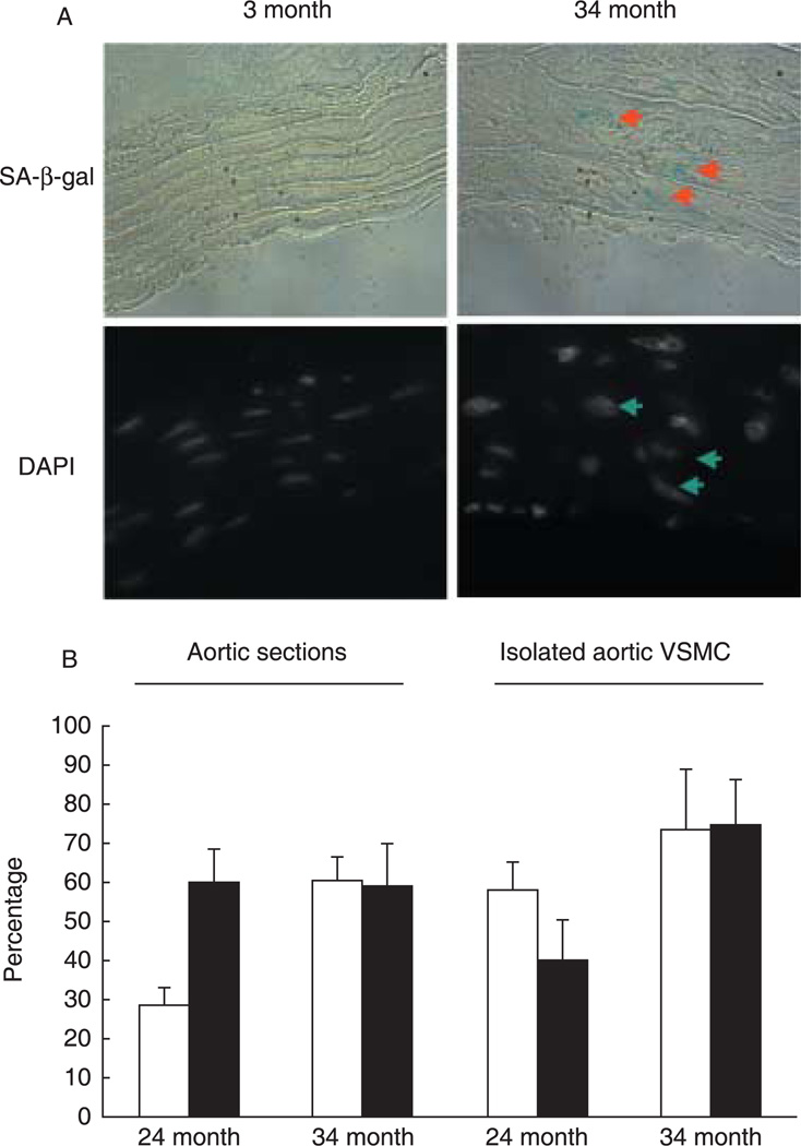
Increased senescence-associated β-galactosidase (SA-β-gal) activity is noted in aortic vascular smooth muscle cells (VSMC) of old rats, and predominantly in the polyploid cells. (A) The SA-β-gal staining assay (blue color; upper panel) was performed on 5-µm cryosectioned aortas and counterstained with 4′,6-diamindino-2-phenylindole (DAPI) to display nuclei (lower panel) (see Supplementary material). The upper panels show aortic sections viewed with phase-contrast microscopy and the bottom panels display respective cell nuclei stained with DAPI (examined with Olympus microscope; ×100 objective). Arrows point to SA-β-gal–positive cells (upper panels) and to corresponding large nuclei (lower panels). (B) Tissue sections or isolated VSMC prepared from 24- or 34-month-old Brown Norway rats as described in the Supplementary material were quantitated. The empty bars depict the percentage of polyploid cells identified based on nuclear size (for tissue sections) or DAPI staining (for isolated VSMC), and the filled bars represent the percentage of polyploid cells that are also SA-β-gal-positive. Three to five slides per rat were analyzed, and a total of two to three rats were examined in each age group. Shown are average percentages ± standard deviations.

