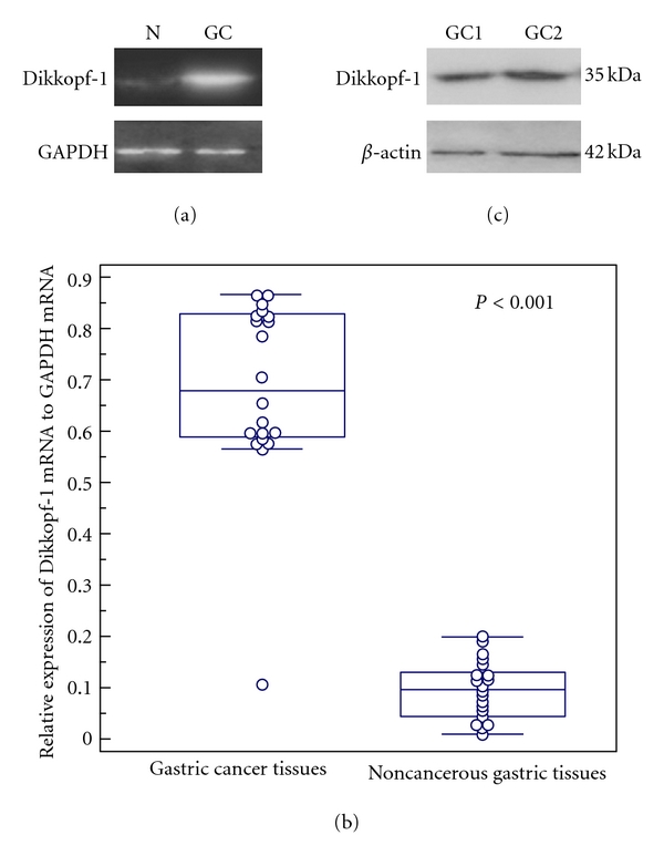
Figure 1.

Real-time quantitative RT-PCR analysis of Dickkopf-1 mRNA expression in 20 pairs of human gastric cancer and adjacent noncancerous human gastric tissues. (a) Gel images of electrophoresis. “N” refers to noncancerous gastric tissues; “GC” refers to gastric cancer tissues. (b) The average level of Dikkopf-1 mRNA expression in gastric cancer tissues was significantly higher than that in noncancerous gastric tissues (P < 0.001). GAPDH gene was used as an internal control. (c) The specificity of Dikkopf-1 antibody was analyzed by western blot testing. A single band of 35 kDa was detected in gastric cancer tissues. β-actin antibody was used as control with the single band of 42 kDa.
