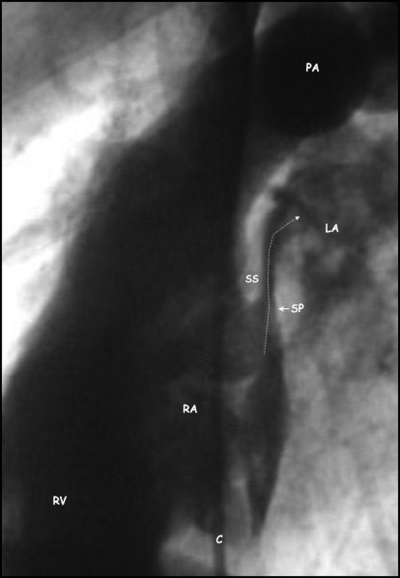
Figure 2.
Angiographic lateral view of a patent foramen ovale showing contrast medium injected into the right atrium (RA) passing (arrow) through the patent foramen ovale between the thick septum secundum (SS) and the thin mobile septum primum (SP) into the left atrium (LA). C, catheter; PA, pulmonary artery; RV, right ventricle.

