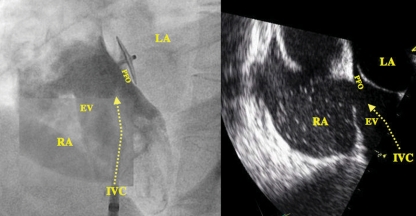
Figure 5.
Angiographic (left panel) and transoesophageal view (right panel) of a patent foramen ovale (PFO) associated with an Eustachian valve (EV) channelling the inflow from the inferior vena cava (IVC) directly onto the PFO. The left panel shows a catheter in the IVC and a 25 mm Amplatzer PFO occluder in the PFO. LA, left atrium; RA, right atrium.

