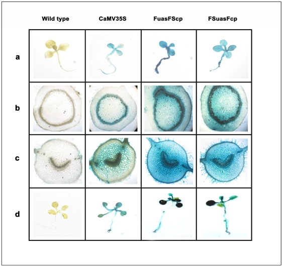
Figure 10. Histochemical localization of GUS activity in transgenic tobacco and Arabidopsis seedlings generated for the respective promoter-GUS constructs.
(a) Histochemical staining of transgenic tobacco seedlings expressing GUS under the control of respective promoter constructs. Photographs were taken using Leica DM LS2 microscope (at 10× magnification) attached to a CCD camera. (b) Histochemical staining of transgenic tobacco stem cross sections expressing GUS under the control of respective promoter constructs. Photographs were taken using Leica DM LS2 microscope (at 10× magnification) attached to a CCD camera. (c) Histochemical staining of transgenic tobacco leaf petiole cross sections expressing GUS under the control of respective promoter constructs. Photographs were taken using Leica DM LS2 microscope (at 10× maginification) attached to a CCD camera. (d) Histochemical staining of transgenic Arabidopsis seedling expressing GUS under the control of respective promoter constructs. Photographs were taken using Leica DM LS2 microscope (at 10× maginification) attached to a CCD camera.

