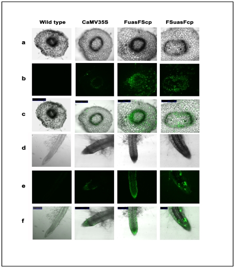
Figure 11. CLSM based analysis of localized GUS expression in transgenic tobacco expressing respective promoter-GUS constructs.
(a) Bright field confocal images of transverse sections of transgenic tobacco stem expressing GUS under the control respective promoter constructs. (b) Fluorescence images of transverse sections of transgenic tobacco stem expressing GUS under the control of respective promoter constructs. (c) Superimposed (bright field and fluorescent) images of transverse sections of tobacco stem expressing GUS under the control of respective promoter constructs. (d) Bright field confocal images of transgenic tobacco root expressing GUS under the control of respective promoter constructs. (e) Fluorescence images of tobacco root expressing GUS under the control of respective promoter constructs. (f) Superimposed (bright field and fluorescent) images of tobacco root expressing GUS under the control of respective promoter constructs. Images were captured using CLSM as described in “Materials and Methods”. Plant samples used were grown aseptically under tissue culture conditions. All figures under row a, b and c were presented in 500 micrometer (µm) scale while figures under row d, e and f were presented in 250 micrometer (µm) scale except figures obtained from promoter FSuasFcp [presented using 100 micrometer (µm) scale].

