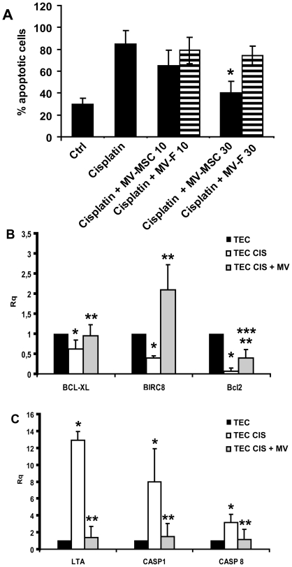
Figure 5. In vitro anti-apoptotic effects of MVs on TECs.
A) The percentage of apoptotic TECs after incubation with 5 µg/ml of cisplatin was evaluated by the Tunel assay. TECs were incubated in the presence of cisplatin with or without different doses of MVs derived from BM-MSCs or fibroblasts (10 or 30 µg/ml) and 3% FCS (Ctrl = TECs incubated 48 hours in the presence of 3% FCS only). Results are expressed as mean±SD of 4 different experiments. Analyses of variance with Newmann-Keuls multicomparison test was performed: *p<0.05 MVs (30 µg) vs vehicle alone. B) Histograms showing the relative expression (Rq) of different anti-apoptotic genes in cisplatin (TEC CIS) and cisplatin-MV treated tubular cells (TEC CIS+MV) in respect to control cells treated with vehicle alone (TEC). Experiments are performed in triplicate. Data was analysed via a Student’s t test (unpaired, 2-tailed); * p<0.05 TEC CIS vs TEC; ** p<0.05 TEC CIS+MV vs TEC CIS; *** p<0.05 TEC CIS+MV vs TEC. C) Histograms showing the relative expression (Rq) of pro-apoptotic genes in cisplatin (TEC CIS) and cisplatin-MV treated tubular cells (TEC CIS+MV) in respect to control cells (TEC). Experiments are performed in triplicate. Data was analysed via a Student’s t test (unpaired, 2-tailed); * p<0.05 TEC CIS vs TEC; ** p<0.05 TEC CIS+MV vs TEC CIS.

