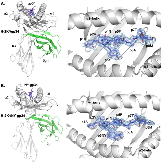
Figure 1. Crystal structures of H-2Kb in complex with gp34 and NY-gp34.
Overall schematic views of the H-2Kb/gp34 and H-2Kb/NY-gp34 MHC complexes are presented in the left part of panels A and B. The α1, α2 and α3 domains of the MHC heavy chain are colored in white. The β2m subunit is colored in green. The peptides are in blue. The 2Fo-Fc electron density maps for the peptides gp34 and NY-gp34 when bound to H-2Kb presented in the right part of panels A and B, respectively, are contoured at 1.0 σ. The final models are displayed for comparison. The peptides, depicted with their N-termini to the left and their C-termini to the right, are displayed ‘from above’ as seen by the TCRs.

