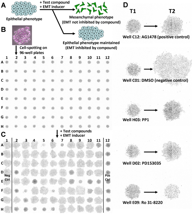
Figure 1. EMT spot migration screening assay overview.
(A) Schematic of the spot migration screening assay to identify EMT inhibitory compounds. EMT can be initiated and maintained in epithelial cells via growth factor signaling. This assay measures the dispersion of cells in the presence of a test compound and an EMT inducer (EGF, HGF or IGF-1). The prevention of cell dispersion directly correlates to the propensity of a test compound to block an induced EMT signaling pathway. (B) Screening assay image acquisition workflow. Robot-assisted plating of H2B-mcherry transfected NBT-II cells into the well centers of 96-well plates. The initial plate image acquired at T1 served as the baseline reference for calculating the CCR and CDR values for each well. The cells were treated with test compounds overnight and further incubated for 24 h with a growth factor to induce EMT. (C) Final plate image acquired at T2 depicted the dispersion response of cells 24 h after addition of the compounds and growth factor treatment. In the example shown, columns 2–11 were treated with 80 different test compounds at 6.67 µM and EGF. Column-1 served as negative controls treated with 0.67% DMSO and EGF, while column-12 served as positive controls treated with 6.67 µM AG1478 and EGF. (D) Magnified images of selected wells acquired at T1 and T2. Wells C12, H03 and D02 are examples of cell colonies treated by compounds that inhibited EGF-initiated cell dispersion and did not inhibit cell growth. Well C01 is a cell colony undergoing EGF-induced EMT without any dispersion inhibition. Well E09 is a cell colony treated by a growth inhibitory or toxic compound.

