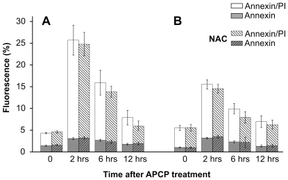
Figure 5. Effects of APCP treatment on externalization of phosphatidylserine.
Cultured human keratocytes (panel A) and conjunctival fibroblasts (panel B) were exposed for 2 minutes to APCP with or without a 5 mM-pretreatment with NAC and subsequently cultured under optimal conditions. The externalization of phosphatidylserine was analyzed by flow cytometry after double staining of the cells with Annexin-V-FITC and propidium iodide, collecting at least 10,000 events. Single or double positive cells are expressed as a percentage of fluorescent intensity. In cells pre-treated with NAC, the increased percentage of AnnexinV/PI positive cells at 2 and 6 hours post-treatment returned to control values after 12 hours. Data are expressed as mean ± SE (error bars) of at least two independent experiments.

