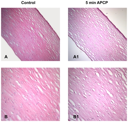
Figure 7. Effects of APCP treatment on ex-vivo human corneas.
The tissue morphology of corneas exposed for 5 minutes to APCP was evaluated. Samples were embedded in paraffin, cut into 5 µm sections, and stained with hematoxylin and eosin. Light microscopic staining was compared to that of unexposed control tissues. A and B: control sections at 100 (A) and 200 (B) magnification; A1 and B1: treated sections at 100 (A1) and 200 (B1) magnification.

