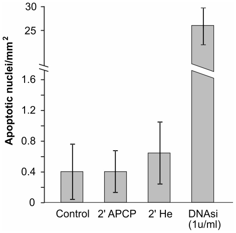
Figure 8. Tunel test on APCP treated corneal tissue.
Apoptotic cells in 5 µm paraffin-embedded corneal tissues exposed for 2 minutes to APCP were identified by labelling DNA strand breaks with biotin-labeled deoxynucleotides and peroxidase. The immunoreaction product was visualized using 3,3′-diaminobenzidine and light microscopy. No significant apoptotic effects were evident in corneal tissues treated with APCP or only with helium (He). Data are expressed as the mean ± SE (error bars) of at least two independent experiments.

