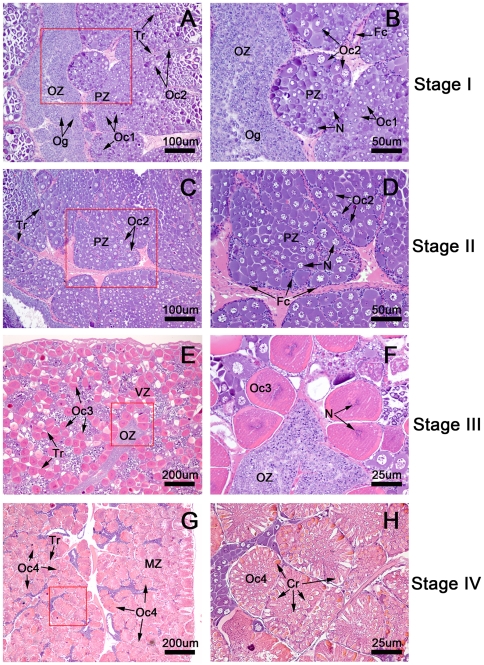
Figure 1. Hematoxylin-and eosin (H&E)-stained paraffin-embedded sections of ovaries at various stages.
The ovaries are composed of lobules separated by connective tissue septae called trabeculae (Tr), with each lobule containing a variety of oocytes surrounded by follicular cells. (A,B) In the stage I ovary (the spent stage), the germ cells present are the oogonia (Og) in the oogenic zone (Oz) and stage 1 oocytes (Oc1) and stage 2 oocytes (Oc2) in the previtellogenic zone (PZ). (C,D) In the stage II ovary (the proliferative stage), the predominant cells are the stage 2 oocytes (Oc2) located in the previtellogenic zone (Pz), while Og and Oc1 are present to a lesser degree. (E,F) In stage III ovaries (the premature stage), the majority of oocytes at stage 3 oocytes (Oc 3) are located in the vitellogenic zone (VZ). Oc3 increase in size during maturation and contain increasing amounts of lipid droplets. The cytoplasm of these oocytes stains pink due to increased eosinophilia. (G,H) In the stage IV ovary (the mature stage), the mature oocytes (Oc4) are the largest and most abundant cells. Oc4 are located in the mature zone (MZ), which occupies almost the entirety of each lobule. The mature oocytes are identifiable by the highly eosinophilic cytoplasm, which is stained a deep pink by the eosin. The cytoplasm of these oocytes contains cortical rods; the oocytes are surrounded by follicular cells (Fc).

