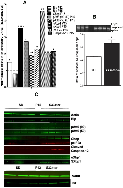
Figure 3. The ER stress markers BIP and CHOP proteins in retinas from S334ter-4 Rho rats.
A: Ratios of normalized S334ter-4 Rho BiP, CHOP, pATF6 (90), pATF6 (50), peIF2α ,spliced Xbp1(sXbp1), unsliced Xbp1 (uXbp1), cleaved caspase-12 proteins to corresponding proteins in SD CHOP were used to register the alteration of protein expression. Normalization of all proteins was done by detecting β-Actin protein The ER stress markers BIP protein was a 1.5-fold upregulated in P12 S334ter-4 Rho retinas compared with SD retinas (0.047±0.008 vs. 0.071±0.005, respectively; P = 0.01). In P15, the level of BiP protein was significantly reduced and was not distinguishable from SD. The Chop protein was also dramatically over-expressed (3.5-fold) (0.016±0.005 in SD vs. 0.60±0.002 S334ter-4 Rho, P = 0.0003) on P15 in S334ter-4 Rho retinas. The full length of pAtf6 protein (90 kD) (the Atf6 pathway) was significantly elevated in S334ter-4 Rho retina by 2.7-fold (0.0017±0.0011 in SD vs. 0.0048±0.0007 in S334ter-4 Rho, P = 0.04). The cleaved pAtf6 (50) was significantly elevated by 1.93-fold inS334ter-4 Rho retina (0.18±0.004 in SD vs. 0.035±0.002, P = 0.004). The peIF2α protein was also significantly increased in S334ter-4 Rho retina and was 0.001±0.0005 in S334ter-4 Rho vs 0.002±0.0004, P = 0.0009. The full length of pAtf6 protein (90 kD) (the Atf6 pathway) was significantly elevated in S334ter-4 Rho retina by 2.7-fold (0.0017±0.0011 in SD vs. 0.0048±0.0007 in S334ter-4 Rho, P = 0.04). The N-terminal of the full-length of Atf6, cleaved pAtf6 was elevated by 1.93-fold and was 0.18±0.004 in SD vs. 0.035±0.002, P = 0.004. We also observed that the peIF2α protein was significantly increased in S334ter-4 Rho retina and was 0.001±0.0005 in S334ter-4 Rho vs 0.002±0.0004, P = 0.0009. The spliced Xbp1 protein was detected in S334ter-4 retina. Its level was a 4.5-fold higher in transgenic retina compared to SD and was 0.022±0.003 in SD and 0.1±0.01, P = 0.001 in S334ter-4 rats. Unspliced Xbp1 protein was increased to a lesser extent (2.3-fold) in S334ter-4 rats and was 0.049±0.008 in SD and 0.13±0.001, P = 0.034 in S334ter-4. Increase in active caspase-12 was observed in S334ter-4 Rho retina. The level of cleaved caspase-12 (20 kD) was elevated in S334ter-4 Rho rats on P15 compared to control over 2-fold and was 0.05±0.007 in SD vs. 0.12±0.02 in S334ter-4 Rho, P = 0.014 on P15. B: Upper panel: Quantification of spliced form of the Xbp1 mRNA (the IRE signaling) detected by RT-PCR reaction. We observed 1.45-fold increased in the spliced form of Xbp1 mRNA in S334ter-4 Rho retina. The ratio of spliced Xbp1 to normalized unspliced Xbp1 mRNA was 0.22±0.0006 in SD vs. 0.033±0.031 in S334ter-4 Rho retina, P = 0.025). Image of the agarose gel loaded with RT-PCR product obtained with Xbp1 specific primers is shown in a lower panel. C: Images of western blots treated with anti-Actin, Bip, CHOP, peIf2α, pATF6, Xbp1 antibodies and detected with secondary antibodies and infrared imaging scanner are presented.

