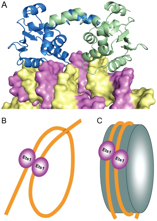
Figure 5. Models of Ets1 binding to widely separated EBS.
(A) Docking of Ets1 homodimer to nucleosomal DNA based on the superimposition of DNA in the (Ets1)2•2DNA structure and the high-resolution structure of a nucleosome core particle (PDB access code 1kx5) [49]. Ets1 molecules are displayed as blue and green cartoons and DNA is displayed as a surface with the strands highlighted in yellow and magenta colors. (B) and (C) Schematic representation of two models of Ets1 cooperative binding to widely separated EBS on promoter DNA: (B) binding via looping of promoter DNA and (C) binding to a nucleosome core particle.

