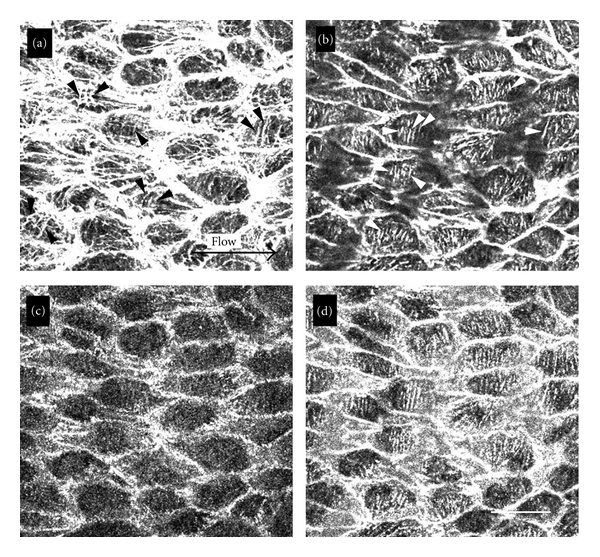
Figure 1.

En face preparations of the renal artery were double-stained with rhodamine-labeled phalloidin (a and b) and antivinculin antibody (c and d). Focus was adjusted to apical (a and c) and basal (b and d) portions of the endothelial cells. In the apical portion of the cells, several stress fibers running perpendicular to the direction of flow were observed (a: arrowheads) among the stress fibers running parallel to the direction of flow (a: arrow). Only stress fibers running perpendicular to the direction of blood flow were seen in the basal portion of the cells (b: arrowheads). All spots showing positive labeling with antivinculin antibody were colocalized with stress fibers (d). Bar: 100 μm.
