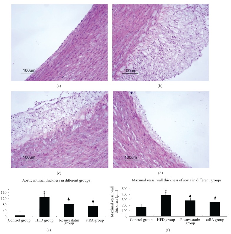
Figure 1.
Characteristics of arterial lesions. Pathological sections of arterial lesions were examined by HE staining in different groups. In the control group (a), the vessel walls were thin and smooth with even thicknesses; in the HFD group (b), there were more foam cells and necrotic substances in the intima. Compared with the HFD group, there were less foam cells, necrotic substances in the intima from rosuvastatin group (c) and atRA group (d) (magnification ×200). Statistical results of aortic intimal thickness (e) and maximal vessel wall thickness (f) among different groups; *P < 0.05 compared to control group, ▲P < 0.05 compared to HFD group.

