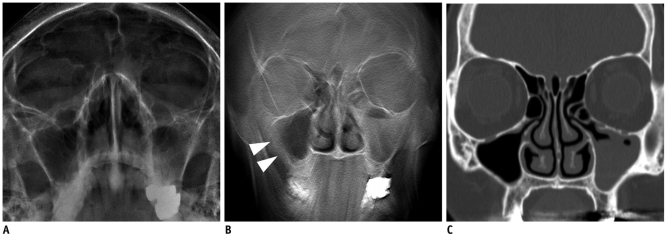
Fig. 4.
Images from 68-year-old woman with cough.
A. Water's view radiograph shows subtle fluid filled left maxillary sinus. There is no motion artifact. B. Tomosynthesis image shows image blurring in right maxillary sinus (arrowheads) which mimic mucoperiosteal thickening. Note prominent motion artifact in mandible area. C. CT image confirms presence of left maxillary sinusitis. Note that right maxillary sinus is normal.

