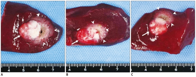
Fig. 2.
Triphenyltetrazolium chloride stained specimens. With triphenyltetrazolium chloride staining, viable tissue with intact mitochondrial enzyme activity stains red, while ablated tissue remains white. Arrow indicating VX2 carcinoma and arrowheads point to ablated area. Ablated area of specimen from group A is larger than that from group B and C.
A. Specimen from group A administered through hepatic artery. B. Specimen from group B administered through auricular vein. C. Specimen from group C, control group, administered phosphate-buffered saline through auricular vein.

