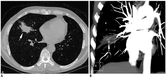
Fig. 1.
Fifty-six-year-old man who had previously received antitubercular treatment, presented with recurrent episodes of hemoptysis and low grade fever.
A. Axial lung window CT image showed nodular lesion in right middle lobe with irregular margins (arrow). B. Coronal maximum-intensity-projection imaging after contrast injection shows multiple hypertrophied arteries and aneurysm (arrow) inside lesion. As result of these findings, biopsy was not attempted. Patient responded to empiric antitubercular treatment and final diagnosis of reactivation tuberculosis was made.

