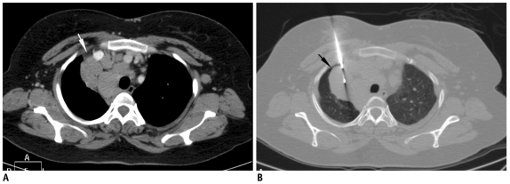
Fig. 19.
Inadvertent pneumothorax during mediastinal biopsy.
A. Localization contrast-enhanced CT scan of 40-year-old woman with multiple mediastinal nodes. Safe window for biopsy was present lateral to right brachiocephalic vein through mediastinal fat (white arrow). B. CT image after advancing needle showed air (black arrow) in path of needle with separation of mediastinal fat from anterior chest wall. Pneumothorax had occurred, either due to inadvertent transgression of interposed parietal pleura or variable respiration bringing aerated lung in path of needle.

