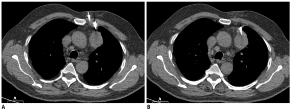
Fig. 26.
Lung biopsy through non-aerated route using mediastinal fat as window.
A. Axial CT image of 68-year-old man shows left upper lobe mass which was abutting mediastinal fat. Biopsy needle was advanced from left parasternal approach lateral to internal thoracic vessels (arrow) using mediastinal fat as window. B. CT image at caudal level showed needle entering into mass without transgression of aerated lung. Biopsy results were suggestive of adenocarcinoma.

