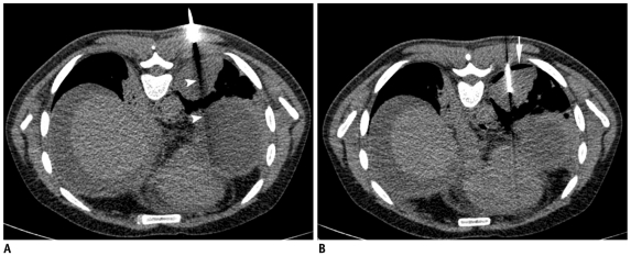
Fig. 28.
Detection of early pneumothorax.
A. Prone CT scan of 32-year-old man with left lower lobe mass and peritoneal deposits. Guiding needle was positioned in chest wall and beam hardening artifact (arrowheads) showed needle trajectory. B. After needle was advanced into mass, air crescent (arrow) was noted separating lung mass from chest wall. This finding was suggestive of development of early pneumothorax and was confirmed on lung window images. Biopsy was quickly completed and patient was placed in "biopsy side down" position. Biopsy results revealed adenocarcinoma.

