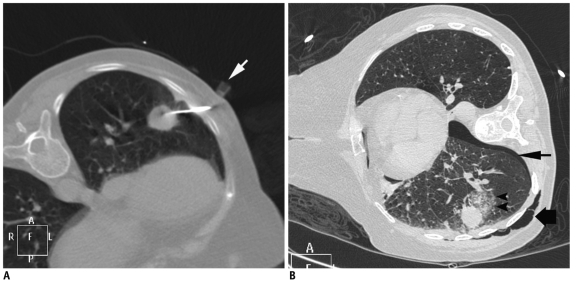
Fig. 29.
Pneumothorax, surgical emphysema and alveolar hemorrhage following lung lesion biopsy.
A. Axial CT image of 73-year-old woman showed left lower lobe nodule. Plastic marker (arrow) was placed close to skin entry site. Due to excessive movement with respiration, lesion could be targeted after puncturing lung twice. B. Post biopsy image showed development of pneumothorax (thin arrow) and surgical emphysema of chest wall (thick arrow). Alveolar hemorrhage is also noted around lesion (arrowheads). Biopsy was suggestive of adenocarcinoma.

