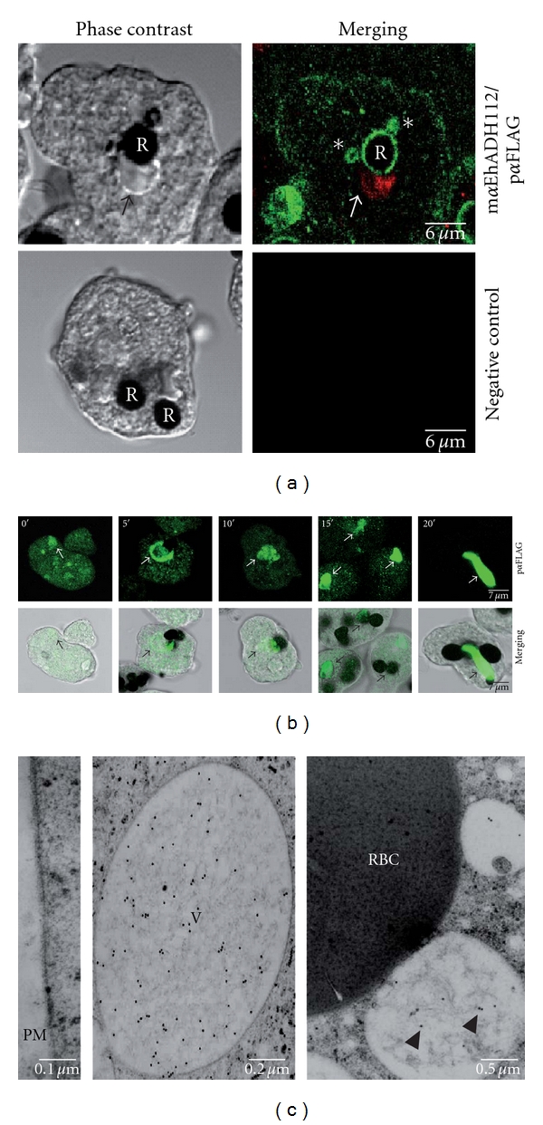
Figure 4.

Differential location of endogenous EhADH112 and the Bro1 recombinant polypeptide in ANeoBro1 trophozoites during phagocytosis. (a) Cellular immunolocation of endogenous EhADH112 and exogenous Bro1 in ANeoBro1 trophozoites after RBC ingestion. Trophozoites were incubated with RBC for 5 min and permeabilized. Top: after RBC contrasting with DAB, EhADH112 was detected by mαEhADH112 antibodies and FITC-labeled anti-mouse secondary antibodies. The FLAG-tagged Bro1 recombinant polypeptide was detected by pαFLAG antibodies and TRITC-labeled anti-rabbit secondary antibodies. Bottom: trophozoites incubated with FITC-labeled anti-mouse IgM and TRITC-labeled anti-rabbit IgG secondary antibodies. Preparations were examined through laser confocal microscopy. R: RBC. Arrows: recombinant Bro1 containing vacuoles. Asterisks: EhADH112 present in endosomal or lysosomal-like compartments. (b) Immunolocation of exogenous Bro1 in ANeoBro1 trophozoites at different times of erythrophagocytosis. Trophozoites were incubated with fresh RBC and treated as described above. The Bro1 recombinant polypeptide was detected by pαFLAG and FITC-labeled anti-rabbit secondary antibodies. Preparations were examined through a laser confocal microscope. Top: confocal sections. Bottom: merging of phase contrast images and laser confocal corresponding sections. Arrows: vesicles and large vacuoles containing exogenous Bro1. (c) Ultrastructural location of exogenous Bro1 in ANeoBro1 trophozoites after RBC ingestion. Ultrathin sections of ANeoBro1 trophozoites were processed for immunogold labeling and TEM as described above. The Bro1 recombinant polypeptide was detected by mαFLAG antibodies and gold-labeled secondary antibodies. Left: plasma membrane (PM). Middle: a huge vacuole. Right: vacuoles in the proximity of RBC after 15 min phagocytosis. V: vacuole. Arrowheads: Bro1 gold-labeled particles.
