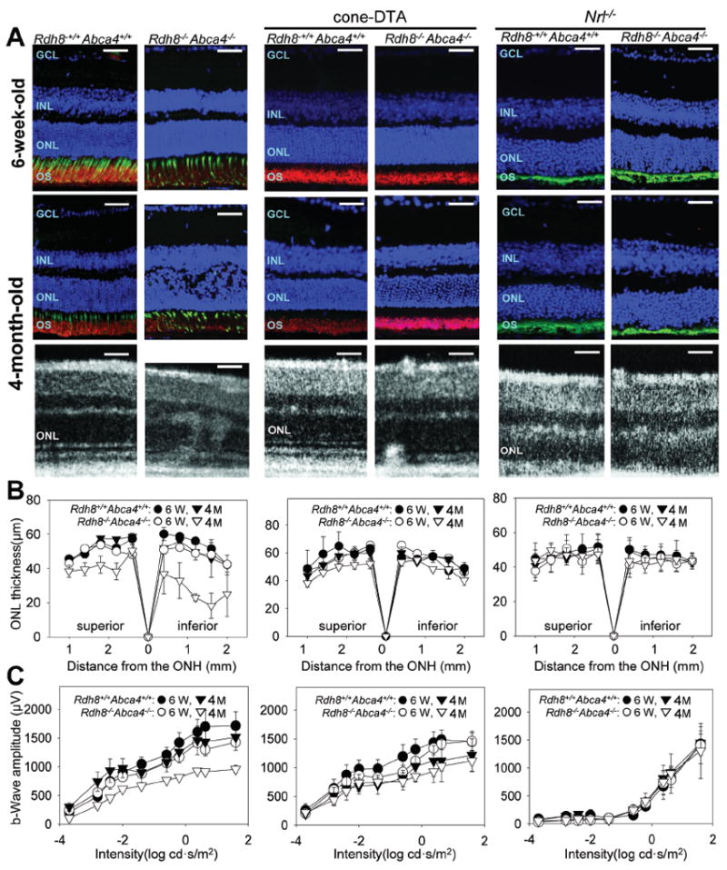
Figure 2. Cone–DTA Rdh8−/− Abca4−/− mice show earlier progression of photoreceptor cell death than Nrl−/− Rdh8−/− Abca4−/− mice, but Rdh8-/- Abca4-/- mice exhibit the earliest and most rapid progression of photoreceptor cell death.

Progression of retinal degeneration in Rdh8−/− Abca4−/− (cone and rod), cone–DTA Rdh8−/− Abca4−/− (rod–only) and Nrl−/− Rdh8−/− Abca4−/− (cone–like–only) mice maintained under a 12 h light (~50 lux) /12 h dark cycle, was evaluated with IHC and SD–OCT imaging (A), measurements of ONL thickness in the superior and inferior retina (B), and scotopic single–flash ERG recordings (C). Cryo sections from 6–week–old and 4–month–old mice were stained with anti–rhodopsin antibody (1D4, red), and PNA (green) and DAPI (blue)(A top and middle panels). Mild photoreceptor cell death was observed in cone–DTA Rdh8−/− Abca4−/− mice whereas no significant changes in ONL thickness were noted in Nrl−/− Rdh8−/− Abca4−/− mice at 4 months of age. However, Rdh8−/− Abca4−/− mice showed significant progression of degenerative changes with disruption of the OS and the ONL at 4 months of age. Similar pathological changes were observed in SD–OCT images obtained from each mouse model at 4 months of age (A bottom panels). Single flash ERG responses were recorded under scotopic conditions and functional b–wave amplitudes were plotted (C). Retinal dysfunction was observed in cone–DTA Rdh8−/− Abca4−/− mice and Rdh8−/− Abca4−/− mice that reflected their corresponding levels of retinal degeneration (C left and middle panels) whereas no significant dysfunction was recorded in Nrl−/− Rdh8−/− Abca4−/− mice (C right panels). Representative images of immunohistochemistry and SD-OCT were obtained in the inferior retina. GCL, ganglion cell layer; INL, inner nuclear layer; ONL, outer nuclear layer; PR, photoreceptor cells. Bars in A, 20 μm. Error bars indicate SDs in B and SEs in C (n> 4).
