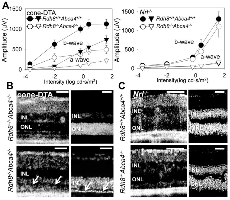
Figure 3. Rod cells are more susceptible to intense light–induced acute degeneration than cone cells.

Six–week–old cone–DTA Rdh8−/− Abca4−/− and Nrl−/− Rdh8−/− Abca4−/− mice were exposed to intense light (10,000 lux) for 30 min to induce acute damage to the retina. Seven days after the light exposure, retinal function and morphology was evaluated by scotopic-ERG recordings and SD–OCT and IHC imaging with DAPI staining. ERG responses in cone-DTARdh8−/− Abca4 −/− mice evidenced significant retinal dysfunction (>60% decreased response) whereas only mild dysfunction (<15% b–wave response depletion at high–intensity stimulation) was noted in Nrl-/- Rdh8−/− Abca4−/− mice. (A: filled symbols indicate mice without deletion of Rdh8 and Abca4; unfilled symbols, Rdh8−/− Abca4−/−; circles, b–waves; triangles, a–waves). Bars indicate SE of means (n > 3). Retinal morphological changes were examined with SD–OCT and IHC with DAPI staining in lesions at superior retina at 500 μm away from the optic nerve head center (B and C). Severe photoreceptor cell death was observed in cone–DTA Rdh8−/− Abca4−/− mice by SD–OCT (B left panel) as manifested by only 1or 2 rows of nuclei in the ONL (white arrows in B right panel). There was no significant morphological change in Nrl−/− Rdh8−/− Abca4−/− mice in SD–OCT (C left panel) and IHC images (C right panel). Error bars indicate SEs in A (n >3). Representative images of SD-OCT were obtained in the inferior retina. ONL, outer nuclear layer. Bars in B, C: 40 μm.
