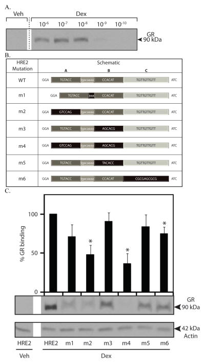
Figure 4.
Dexamethasone-dependent binding of GR to the Edn1 HRE2. A. GR binding to the Edn1 HRE2 region was evaluated with DNA affinity purification assays (DAPA) performed on mIMCD-3 cells treated with varying concentrations of dexamethasone (0.1 nM to 1 μM) for 1 h. B. Mutations to the Edn1 HRE2 DAPA probes. Three putative receptor binding half-sites are indicated as A, B and C. The spacer region is shown in light grey with white text. Mutated regions are highlighted in black. C. DAPAs were performed using HRE2 mutant probes on mIMCD-3 cells treated with 1 μM dexamethasone for 1 h. Densitometric values are indicated above each band and are shown as percent (%) GR binding. GR binding to the HRE2 wild-type probe in the presence of dexamethasone is set to 100%. (*p < 0.05, n ≥ 3)

