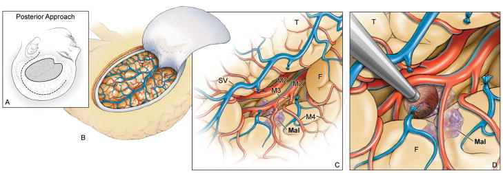
Figure 3.
(A) With the posterior transsylvian-transinsular approach, the head is positioned nearly laterally to align the plane of the opercular cleft vertically. A question mark skin incision (dashed line) is used and the pterional craniotomy is extended posteriorly to incorporate more parietal and posterior temporal bone (solid line). (B) The craniotomy exposes the entire Sylvian fissure, from the pterion proximally to the operculum distally. (C) Dissection begins at the same place as with the anterior approach, but proceeds in the opposite direction. Cortical (M4) and opercular arteries (M3) are followed down to the depths of the opercular cleft to reach the long gyri of the insula. (D) Lesions not on the insular surface are accessed through a small cortical incision. Abbreviations: F = frontal lobe; T = temporal lobe; Mal = malformation; SV = Sylvian vein; M2 = insular segment of MCA.

